Label the tooth diagram quiz online
Take an alternative quiz on labelling a tooth below
Label the tooth diagram quiz online. This is a bilogy quiz online for children in 2nd, 3rd, 4th, 5th and 6th grades. In this quiz on tooth anatomy, students will learn how to draw and label a tooth. Each tooth has different parts and each part plays a particular role. This quiz is interactive and feedback in instant. Students can freely practice and home or in school. This is a great free teaching resource for parents and teachers. There is a worksheet below on a tooth diagram to label .
Label Teeth Diagram
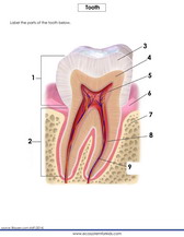
Digestion begins in the mouth. When we ingest food in the mouth, our teeth help us to chew the food into tiny particles that can be further digested when they get into the stomach. The ability of the teeth to chew hard food particles into smaller pieces tells of an organ that is built tough to resist breaking off. The tooth is made up of three distinct parts as follows: the crown, the neck and the root. Each part is further divided into other distinct units with each one displaying a unique characteristic. The crown is made up of the enamel; the white hard part that is visible from the outside. It is what we brush with tooth paste to keep clean and void of bacteria. Directly underneath the enamel is the dentine and underneath the dentin is the pulp cavity. The pulp cavity contains the root canal within which are blood vessels and nerves. The sensitivity of our teeth is accounted for by the presence of these sensory nerves underneath it. The neck is the section that is in contact with the gum. The gum itself is the red or dark part to which our teeth stick. The teeth are actually stuck to this section by what is called the root. Think about the function plant roots play; it is the same function the roots of our teeth play. They stick our teeth right into the bone passing through the gum. The roots are held in place in the gum through what is called cement. The terms used to label a teeth diagram are familiar words you see in other contexts. Check out the worksheet below for more practice on how to label a tool.
Tooth Anatomy: A Comprehensive Guide
We, as dental professionals, believe that understanding the anatomy of teeth is crucial in maintaining good oral health. In this article, we will delve deep into the structure of teeth, their functions, and how they contribute to overall oral health. By the end of this article, you will have a comprehensive understanding of tooth anatomy.
The Crown: The Visible Part of the Tooth
The crown is the visible part of the tooth, and it is covered with enamel, the hardest substance in the human body. Enamel protects the tooth from decay and erosion caused by bacteria and acid. The crown consists of three layers: enamel, dentin, and pulp. Enamel is the outermost layer, and it is white in color. Dentin is the layer beneath enamel, and it is yellowish in color. Pulp is the innermost layer, and it contains nerves and blood vessels.
The Root: The Anchor of the Tooth
The root is the part of the tooth that is embedded in the jawbone. It is covered with cementum, a layer that attaches the tooth to the bone. The root also has three layers: cementum, dentin, and pulp. The pulp in the root is responsible for nourishing the tooth.
The Periodontium: The Support System of the Tooth
The periodontium is the supporting structure of the tooth. It consists of four parts: the gingiva (gums), the periodontal ligament, cementum, and alveolar bone. The gingiva is the soft tissue that surrounds the teeth and covers the jawbone. The periodontal ligament is the connective tissue that attaches the tooth to the bone. Cementum covers the root, and the alveolar bone is the bone that supports the teeth.
Functions of Teeth
Teeth have several functions, and each type of tooth has a specific purpose. Incisors are the teeth in the front of the mouth, and they are used for cutting and biting. Canines are the pointed teeth next to the incisors, and they are used for tearing and gripping food. Premolars are located between the canines and molars, and they are used for grinding and crushing food. Molars are the largest teeth in the back of the mouth, and they are also used for grinding and crushing food.
The Importance of Maintaining Good Oral Health
Maintaining good oral health is crucial for overall health and well-being. Poor oral health can lead to several dental problems such as cavities, gum disease, and tooth loss. It can also lead to other health problems such as heart disease, diabetes, and respiratory infections.
Preventing dental problems requires a daily oral hygiene routine. Brushing twice a day with a fluoride toothpaste, flossing daily, and visiting the dentist regularly for checkups and cleanings are essential for maintaining good oral health.
In Conclusion
Understanding the anatomy of teeth is essential in maintaining good oral health. Teeth are complex structures that serve several functions. The crown is the visible part of the tooth, the root anchors the tooth to the jawbone, and the periodontium is the support system of the tooth. Maintaining good oral health requires a daily oral hygiene routine and regular visits to the dentist.
We hope this comprehensive guide to tooth anatomy has been informative and has provided you with a better understanding of the importance of good oral health. By taking care of your teeth, you can enjoy a healthy and happy smile for years to come.
FAQ:
-
Why is a labelled tooth diagram important?
A labelled tooth diagram is important because it provides a clear and concise representation of the human dentition, allowing dental professionals and students to easily understand the location and anatomy of each tooth in the mouth. It is also a valuable tool for communication between dental professionals and patients, as it allows dentists to explain dental problems and procedures in a clear and understandable manner.
-
How are teeth represented in a labelled tooth diagram?
Teeth are represented in a labelled tooth diagram by a standardized symbol that corresponds to their anatomy. Incisors are represented by a square symbol, canines by a triangle, premolars by a diamond, and molars by a circle. The symbols are also color-coded to indicate the different quadrants of the mouth.
-
What are the functions of each type of tooth?
Incisors are located at the front of the mouth and are used for biting and cutting food. Canines are located next to the incisors and are pointed teeth that are used for tearing and shredding food. Premolars and molars are located in the back of the mouth and are used for grinding and crushing food.
-
How is a labelled tooth diagram used in dental education?
A labelled tooth diagram is a useful tool in dental education because it allows students to visualize the location of each tooth in the mouth and to understand its unique features. It is also a useful reference for students when studying dental anatomy and for dental professionals when explaining dental problems and procedures to patients.
-
How is a labelled tooth diagram used in communication between dental professionals and patients?
A labelled tooth diagram is a valuable tool for communication between dental professionals and patients because it allows dentists to explain the location and anatomy of dental problems or procedures to their patients in a clear and understandable manner. Patients can also use the diagram to better understand their dental condition and the recommended treatment plan.